تعريف
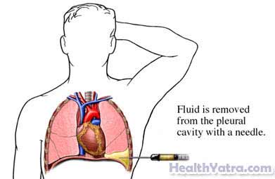
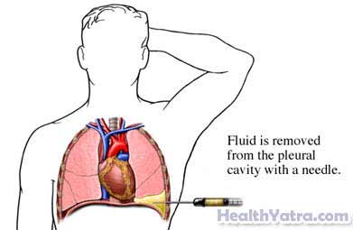
A pleural effusion is a build-up of fluid in the space between the lungs and the chest wall. This space is called the pleural space. Thoracentesis is a procedure to remove fluid from this area.
There are two types of thoracentesis:
- Therapeutic thoracentesis—to relieve the symptoms of fluid accumulation
- Diagnostic thoracentesis—to test for the cause of the fluid build-up
أسباب هذا الإجراء
There is always a small amount of fluid in the pleural space. The fluid helps to lubricate the area. When too much fluid builds up in this space, it can make it difficult to breathe.
Your doctor may want to test some of the fluid after removing it. The build-up of fluid can be a symptom of diseases or disorders, such as:
- Congestive heart failure (CHF)
- Lung infections
- مرض كلوي
- Pulmonary embolism —a clot that travels to the lung
- Cancer
- مرض الكبد
- التهاب البنكرياس
Smoking may increase the risk of complications.
المضاعفات المحتملة
Complications are rare, but no procedure is completely free of risk. If you are planning to have a thoracentesis, your سيقوم الطبيب بمراجعة القائمة من المضاعفات المحتملة، والتي قد تشمل:
- A collapsed lung
- Fluid building up again
- نزيف
- العدوى
- Damage to the liver or spleen
تشمل العوامل التي قد تزيد من خطر حدوث مضاعفات ما يلي:
- A history of lung surgery
- A long-term, irreversible lung disease, such as emphysema or asthma
- Anything affecting normal blood clotting
ما يمكن توقعه
قبل الإجراء
Your doctor may order:
- A complete physical exam
- الأشعة السينية
- الاشعة المقطعية
- الموجات فوق الصوتية
- تحاليل الدم
التخدير
A local anesthetic will be used. It will numb the area where the needle will be inserted.
وصف الإجراء
You may be asked to sit upright on the edge of a bed or chair. Your arms will be resting on a nearby table. If your procedure involves a CT scan, you may be asked to lie on a table. Try to avoid coughing, breathing deeply, or moving during the procedure.
A small patch of skin on your back, chest, or under your armpit will be sterilized. Anesthesia will be applied to this patch. It will help numb the area.
The doctor may use ultrasound or CT scan images. These images will help guide the needle and monitor the fluid. A needle or thin plastic catheter will be inserted between your ribs. The needle or catheter is then passed into the pleural space. Some or all of the fluid will be drawn into the syringe.

كم من الوقت سيستغرق ؟
About 15 minutes
هل ستسبب الالم؟
You may feel slight pain or a stinging when the needle is first inserted. As the fluid is being extracted, you may feel a sense of pulling. Tell your doctor or nurse if you feel extreme pain, any shortness of breath, or faint.
رعاية ما بعد العملية
في مركز الرعاية
If the thoracentesis is being done for diagnostic reasons, the fluid will be sent to a lab for testing. Often, another chest x-ray will be done to ensure that the fluid has been removed and that there is no sign of a collapsed lung.
في البيت
Keep the area of skin where the needle was inserted clean and dry. To help make your recovery smooth, be sure to follow your doctor’s instructions .
If a diagnostic thoracentesis was done, ask your doctor when to expect the results.
استدعاء الطبيب
بعد وصوله الى المنزل ، اتصل بطبيبك إذا كان أي من الحالات التالية:
- علامات الإصابة, بما في ذلك حمى وقشعريرة
- Redness, swelling, increasing pain, excessive bleeding, or any discharge from the insertion site
- ألم لا يمكنك السيطرة عليه بالأدوية التي أعطيت لك
- سعال أو ضيق في التنفس أو ألم في الصدر
- سعال الدم
- Pain when taking a deep breath
إذا كنت تعتقد أن لديك حالة طارئة ، فاتصل بالمساعدة الطبية على الفور.

